Your cart is empty.
Tecan uses cookies to improve our website. By continuing to browse our website, you accept our cookie policy.
Cell counting, viability analysis and live-cell imaging in a multimode reader? With Tecan’s Spark®, all this is possible. Spark has a simple and cost-effective cell imaging module built directly into the reader, enabling automated cell counting and viability analysis in slides, as well as bright field imaging and automated confluence assessment in microplates. With these integrated features, Spark delivers better quality and more relevant data for your cell-based research and opens the door to the exciting world of live-cell imaging.
***************************************************************************************
***************************************************************************************
Tab 01 / Live viewer
***************************************************************************************
***************************************************************************************
The ultimate check before you start your assays.
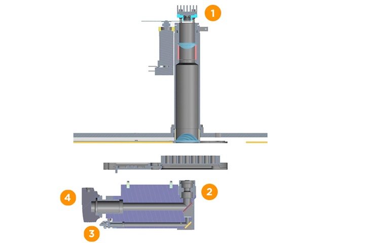
With Spark, you can turn your microplate reader into an automated microscope at the touch of a button. Spark’s imaging module incudes LED bright field illumination, 4x objective magnification, a CMOS 1.3 megapixel camera and a robust laser-based autofocus system – delivering performance to rival an automated microscope.
The SparkControl™ software's Live Viewer application allows the user to automatically acquire and instantly view images of defined positions in a microplate well or Tecan's proprietary Cell Chip™.
Easy intra- and inter-well navigation allows straightforward bright field microscopy within the reader.
Live Viewer also allows the user to manually re-adjust the focus offset before starting the automated well imaging method.
***************************************************************************************
***************************************************************************************
Tab 02 / Cell counting and viability
***************************************************************************************
***************************************************************************************
Counting and viability assessment of cells in solution, such as trypsinized cells, is a daily task in any cell culture lab. Spark’s cell imaging module offers automated, label-free cell counting and trypan blue-based cell viability analysis with an easy-to-use, disposable Cell Chip™.
Each Cell Chip has two sample chambers which can accommodate independent cell suspensions or replicates of the same sample. Four Cell Chips can be loaded onto the Cell Chip adapter, allowing eight samples to be analyzed in less than two minutes.
For cell viability assessments, the cell suspension can be quickly and easily stained with trypan blue to discriminate between live and dead cells.
The cell counting and cell viability apps both offer the same features, the only difference is that the viability app automatically takes into account a 1:1 trypan blue dilution when calculating the results.
Users can set the cell size to between 4 and 90 µm before starting the measurement, allowing you to tailor the assay to your chosen cell line.
For a better counting accuracy and reproducibility, the app also allows you to select one, four or eight images per chamber. This enables fast cell counting for applications with relatively high cell concentrations – around 1x10⁶ cells/ml – by taking a single image at the center of each chamber. In contrast, for low cell concentrations of less than 1x10⁴ cells/ml, eight separate images can be used per sample to dramatically improve accuracy for cell concentration measurements. Furthermore, the two chambers of each slide can be defined as 'duplicates' for automatic evaluation.
Cell counting and cell viability results are clearly presented in a table and histogram on the software dashboard, allowing straightforward size distribution analysis of the cell population. All results are then saved in a PDF report for easy and convenient data storage.
This Technical Note describes the functions and benefits of the Spark multimode microplate reader and cell imaging module, as well as the SparkControl software and its dedicated Cell Counting and Cell Viability Application.
In this technical note, we demonstrate the superior performance of the Spark reader compared to two other multimode readers from different providers. The detection limit for the far red fluorescent dye Alexa Fluor 647 – which has a fluorescence spectrum comparable to Cy5 – was determined, operating each instrument in MCR mode.
Cell counting, viability analysis and determination of size distribution are routinely performed by researchers across multiple disciplines, from standard cell passaging for downstream experiments to using insect cells infected with baculovirus to produce recombinant proteins, or determining the differentiation capacity of stem cells. Tecan’s Spark multimode reader has an integrated, bright field cell imaging module. This patent-pending system enables label-free cell counting, size distribution determination and trypan blue-based cell viability analysis with an easy-to-use, disposable Cell Chip™. In this study, the analysis of cell concentration, cell viability and size distribution for the determination of the optimal protein production rate in recombinant Sf21 cell cultures is presented.
***************************************************************************************
***************************************************************************************
Tab 03 / Live-cell imaging and confluence
***************************************************************************************
***************************************************************************************
The number of cells that adhere to the bottom of a microplate well in a given timeframe can be difficult to quantify, as the growth and adherence rates are influenced by a number of factors. Consistent determination of cell confluence and estimation of cell numbers are therefore important for reproducible assays and interpretation of experimental data.
Spark’s cell imaging module allows you to precisely determine the confluence level in a well from 10 to 90 %, giving you a new level of data quality for your assays.
The easy-to-use SparkControl™ software app allows you to select different imaging areas within the well. Depending on the assay, you can either select just a single image of the well center or a full-well picture composed of multiple images, with automatic well border detection to compensate for variations in microplate dimensions.
Confluence is assessed in real time, with data displayed on the dashboard and exported directly to Excel® for further data analysis. Each confluence value is linked directly to the raw and analyzed images, enabling quick and easy visual confirmation of cell confluence.
Confluence determination is completely independent of cell morphology, using an algorithm designed to offer exceptional sensitivity, even when working with low contrast cells, such as fibroblasts. This makes Spark's confluence measurement app compatible with virtually any cell type, offering an easy-to-use approach to improve your assays.
Sample image of CHO (chinese hamster ovary) cells imaged in a transparent, 96-well microplate.
Sample image of HeLa cells (human epithelial cervix adenocarcinoma) imaged in a transparent, 24-well microplate.
Sample image of A431 (human epidermoid carcinoma) cells imaged in a transparent, 48-well microplate.
Sample image of HepG2 (human liver cancer) cells imaged in a transparent, 12-well microplate.
Sample image of human fibroblasts imaged in a 96-well transparent microplate.
For many cell biology applications – such as the identification and separation of high expression clones or knock-down candidates, and small-scale antibody or protein production – it is important to ensure that cell lines originates from a single cell, i.e. they are monoclonal. This can be a major bottleneck, especially for academic laboratories, as dedicated automated microscopy systems are both very expensive and complex to operate, meaning verification of monoclonality is often performed by manual microscopy.
Tecan’s Spark multimode microplate reader with integrated cell imaging simplifies cell culture, featuring quality checks and signal normalization to cell confluence, thereby boosting automation of cell-based studies.
Cell migration is a central process in the development and maintenance of multicellular organisms, with tissue formation during embryonic development, wound healing and immune responses all requiring the orchestrated movement and assembly of cells.
***************************************************************************************
***************************************************************************************
Tab 04 / Live-cell package
***************************************************************************************
***************************************************************************************
Your life as a cell biologist revolves around keeping your cells happy, no matter the time or day. Take back control of your time and keep your cells happy by automating your workflows. Spark brings together the capabilities of an automated incubator, a microplate reader and a cell imager in one, offering new possibilities for you and your lab.
Spark is equipped with precise temperature control to enable uniform heating of your microplate. To help ensure even temperature distribution, the microplate chamber is thermally isolated from the optics, guaranteeing stable and precise temperature regulation for your assays.
Spark adds another layer of environmental controls by putting CO2/O2 partial pressure control at your fingertips. An integrated Gas Control Module (GCM™) offers CO2 regulation from 0.04 to 10 % (vol.) and O2 from 0.1 to 21 % (vol.), creating an ideal environment for your cells. CO2 regulation maintains cell health during long-term kinetic studies, while O2 control allows you to maintain the correct atmosphere for microaerophilic organisms or mimic physiologic conditions.
Humidity regulation at levels of 95 % or higher is another key element for good cell viability and growth, and minimizing evaporation is essential to maintain constant concentrations during long-term assays. Spark’s Humidity Cassette is a cost-effective solution to minimize evaporation in any SBS-format microplate.
This tool incorporates a filling moat and has a specialized lid for use with our unique Lid Lifter™, giving you passive humidity control for any assay. Simply add ddH2O to the filling moat and you have an instant, no-fuss humidified atmosphere in your microplate chamber.
Spark gives you access to a new type of reader automation – integrated lid lifting – creating new workflow possibilities, reducing the risk of sample contamination and helping to establish an ideal environment for long-term kinetic assays. Whether you want to dispense compounds without the need for manual intervention, or maintain optimal environmental conditions without compromising evaporation protection, Spark is the only reader up to the task.
SparkControl™ combines automation of long-term kinetic assays with advanced features such as kinetic conditioning, remote operation and Open Kinetics, providing a hands-off solution for complex experimental set-ups.
Many live-cell kinetic experiments require specific actions – for example, addition of a compound – once a certain signal level or OD value is reached. Multimode readers equipped with manual environmental controls can make this type of study cumbersome or error-prone.
SparkControl’s kinetic conditioning feature allows you to program the system to automatically perform a required action as soon as a predefined signal is reached in a control well, fitting almost any assay workflow you can think of, in un-lidded or lidded microplates.
SparkControl’s remote control feature allows you to monitor or operate the instrument from outside of the lab. This enables full system control without the need to sit in front of the reader for hours; if you are connected to the internet, then you are connected to your experiment.
A multimode reader is frequently used for multiple applications by various users. Long-term live-cell kinetics assays can block the reader for an entire day, limiting overall lab throughput. SparkControl offers an efficient solution: Open Kinetics. This feature allows you to pause and resume kinetic experiments, enabling additional measurements to be performed throughout long-term studies. This frees up the instrument for other users, eliminating one of the major bottlenecks of shared lab equipment, without compromising your valuable research.
Tecan’s Spark multimode microplate reader was used to identify metabolic changes during the differentiation of human mesenchymal stem cells and to assess and quantify cell viability, cytotoxicity and apoptosis, using several well-established and reliable cell-based assays developed by PromoCell.
Tecan’s Spark multimode microplate reader was used for the real-time analysis of human stem cell proliferation and differentiation, using established assays for cell viability, proliferation and apoptosis assessment in a reliable and convenient manner.
Assaying cells in an appropriate window of time after a compound-¬mediated cytotoxic event can be extremely challenging. This note describes the automation for kinetic monitoring of cytotoxicity and viability using three Promega assays for viability, cytotoxicity and apoptosis assessment with the Spark multimode reader.
***************************************************************************************
***************************************************************************************
Tab 05 / Specification
***************************************************************************************
***************************************************************************************
1 - Imaging mode: Bright field
2 - Objective magnification: 4x
3 - Autofocus: Laser-based
4 - Camera: CMOS 1.3 megapixel
Illumination: High power LED (590 nm)
Optical resolution: >3 µm
Read speed: <5min
(1 image per well for 96-well plate)
For research use only.